


Building AI software for medical imaging isn't just about writing code; it's about creating algorithms that can see what the human eye might miss in X-rays, CT scans, and MRIs. The goal is to provide clinicians with a powerful co-pilot, enabling them to diagnose diseases earlier, faster, and with greater confidence. This is where deep medical insight and sharp data science come together, creating tools that can improve patient outcomes and make hospital workflows a lot smoother. Pulling this off successfully requires a unique mix of clinical expertise, regulatory savvy, and specialised AI development services.
The New Frontier of AI in Medical Diagnostics
What was once a futuristic talking point is now a clinical reality. AI is no longer a "what if" in medical imaging; it's a "how to," and this guide is your roadmap. We're going to walk through how these tools are already helping radiologists improve their diagnostic accuracy, streamline their day, and ultimately, drive better patient care.
In places like Canada, the push for this technology is massive, highlighting a real need for software that fits into the messy, complex world of clinical practice. Building these solutions is more than a technical challenge. It demands a genuine understanding of a radiologist's workflow, strict data privacy rules, and the maze of regulatory approvals. This is why finding the right development partner is so critical; they can help you tackle the big hurdles, from reducing diagnostic errors to securely managing enormous imaging datasets.
A High-Level View of the Development Process
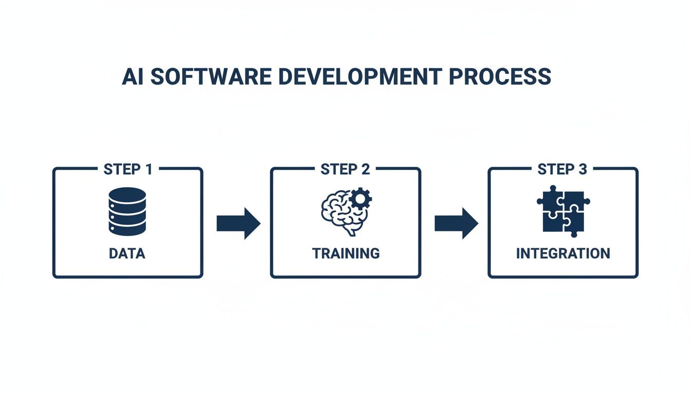
At its heart, the path from a folder of raw medical scans to a trusted clinical tool follows a clear, structured sequence. Think of it as a journey through distinct phases, starting with data, moving to the model, and ending with integration into the clinical environment.
Before we dive deep into each stage, let's get a bird's-eye view of the entire development lifecycle. The table below outlines the key phases, what happens in each one, and what you should have at the end of it.
Key Stages in AI Medical Imaging Software Development
| Phase | Core Activities | Key Outcome |
|---|---|---|
| 1. Data Foundation | Sourcing, collecting, and de-identifying high-quality medical imaging data. Expert annotation and labelling. | A curated, regulatory-compliant, and accurately labelled dataset ready for model training. |
| 2. Model Development | Selecting the right AI architecture (e.g., CNNs), training the model, and iteratively tuning its parameters. | A high-performing AI model that accurately identifies the target features in medical images. |
| 3. Validation & Approval | Rigorous evaluation on unseen data, clinical validation studies, and navigating regulatory pathways (e.g., Health Canada, FDA). | A clinically validated and regulatory-cleared algorithm proven to be safe and effective. |
| 4. System Integration | Building the software architecture to integrate with hospital systems like DICOM and PACS. | A deployable software product that fits seamlessly into existing clinical workflows. |
| 5. Deployment & MLOps | Deploying the model into production, monitoring its performance, and establishing processes for continuous updates. | A live, operational AI tool that delivers consistent value and can be maintained over time. |
This roadmap provides a clear structure for the complex journey ahead. Each step logically builds on the one before it, showing just how crucial a solid foundation is for success.
As you can see, every stage is interconnected. A robust, well-annotated dataset is the bedrock for training a reliable AI model, which in turn must be built to integrate smoothly into a real-world medical setting. It's a process that demands both technical skill and a profound respect for the clinical context where the software will be used. We've previously explored this balance in our overview of AI for medical imaging and diagnostics.
Market Growth and Opportunity
The appetite for these advanced tools is undeniable. Just look at the numbers. In Canada, the AI in medical imaging market is expected to hit USD 367.2 million by 2030, growing at a blistering compound annual growth rate (CAGR) of 34.8%. That's not just growth; it's a clear signal of intense demand for custom AI solutions built for Canadian healthcare providers. To put that in perspective, this pace is well ahead of the global average, marking Canada as a prime market.
This explosive growth tells us one thing: hospitals and clinics aren't just dipping their toes in the water anymore. They are actively investing in AI as a core piece of their strategy for the future of patient care. The real opportunity is for those who can build solutions that are not only technically brilliant but also clinically proven and effortlessly integrated.
This guide will break down the entire process of AI medical imaging software development into clear, actionable steps. From the nitty-gritty of data preparation to the high stakes of regulatory approval, you'll get the practical insights you need to take your idea and turn it into a real, market-ready medical tool.
Building a Solid Foundation with Medical Data
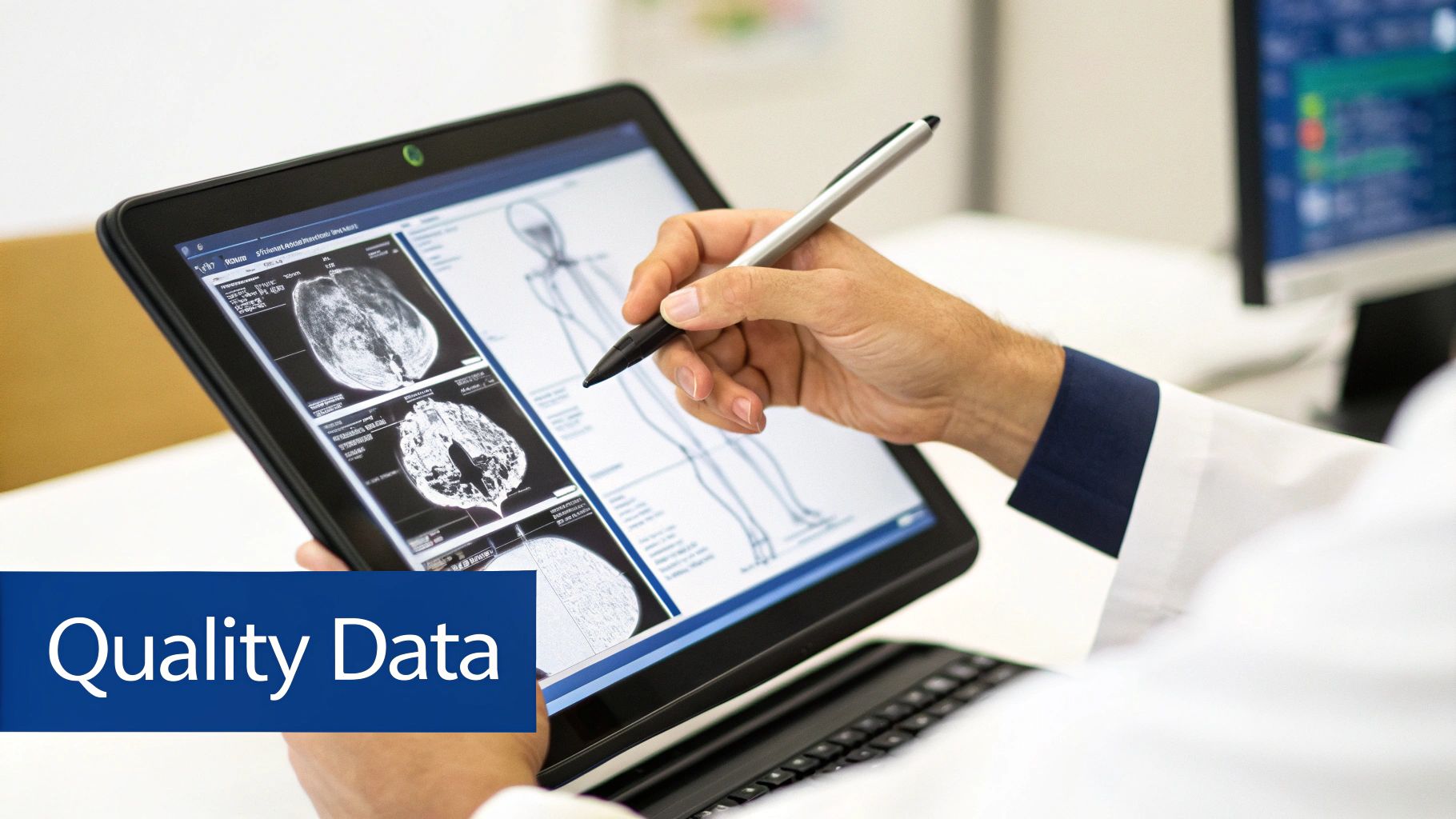
Every powerful AI algorithm starts with one thing: high-quality, relevant data. When you're developing AI medical imaging software, this isn't just a step; it's the entire foundation. The accuracy, reliability, and ultimately, the clinical trustworthiness of your software are a direct reflection of the data you train it on.
Getting your hands on that data is the first challenge. Medical images are often siloed in different Picture Archiving and Communication Systems (PACS), buried in clinical trial databases, or scattered across electronic health records. Pulling these disparate datasets together requires a thoughtful strategy for secure transfer and interoperability.
Sourcing and Privacy Compliance
Before you can even think about training a model, every single image has to be scrubbed of personally identifiable information. This process, known as de-identification, is an absolute must for complying with strict privacy regulations like Canada's Personal Information Protection and Electronic Documents Act (PIPEDA) or HIPAA in the United States.
Failing to properly anonymise data isn't a simple mistake; it's a serious legal and ethical line to cross. A bulletproof de-identification protocol means removing:
Patient names, IDs, and other direct identifiers from DICOM tags.
Any hidden metadata that could accidentally point back to a patient.
"Burnt-in" text on the images themselves that might contain sensitive info.
Navigating these common data integration problems is crucial to building a clean, cohesive dataset from the get-go.
The Art and Science of Annotation
With a clean, compliant dataset in hand, the next phase is annotation. This is where you label the images to teach the AI what to look for, transforming raw pixels into structured, meaningful information. Honestly, this is often the most painstaking and resource-intensive part of the entire project.
The quality of your annotation establishes the "ground truth" for your AI model. If the labels are inconsistent or inaccurate, you're teaching the model the wrong lessons. This leads to unreliable results that are, frankly, useless in a clinical setting.
This isn't a task you can just hand off to anyone. True ground-truth accuracy is only possible when you collaborate closely with board-certified radiologists or other specialists. Their expertise is what guarantees the labels are not just technically correct but also clinically sound. You can learn more about the complexities involved in our guide to healthcare data management software development.
Annotation Techniques in Practice
The right annotation method depends entirely on the clinical question you're trying to answer. The level of detail needed can swing wildly from one project to another, which directly impacts the time, cost, and complexity of the work.
Here are a few common techniques and how they're used in the real world:
Bounding Boxes: This is the simplest approach – drawing a box around a region of interest. It's perfect for straightforward detection tasks, like finding and flagging a suspected tumour on a CT scan.
Semantic Segmentation: This is much more detailed. Here, every single pixel in an image gets assigned a class. For example, in a cardiac MRI, you might segment the left ventricle, right ventricle, and myocardium to measure their volume and function.
Polygonal Segmentation: When a simple box won't cut it, this technique allows annotators to trace the precise outline of an irregular shape, like a complex lesion or a specific organ.
Each technique serves a distinct purpose. Choosing the right one is key to training a model that actually performs the specific task you designed it for. This data pipeline, built on rigorous privacy protocols and expert-led annotation, is what separates a lab prototype from a true, medical-grade AI solution that can stand up to clinical and regulatory scrutiny.
Alright, you've got your pristine, expertly annotated dataset. Now for the exciting part – breathing life into your AI. This is where we turn that curated data into the core intelligence of your software, an algorithm that can make complex clinical judgements. It's a process of picking the right tools for the job, training them meticulously, and then putting them through the wringer with rigorous validation.
The model you choose hinges entirely on the clinical problem you're trying to solve. There's no magic bullet here; different tasks demand different architectural blueprints.
Choosing the Right Model Architecture
In the world of medical imaging AI, two architectures have become the go-to workhorses: Convolutional Neural Networks (CNNs) and U-Nets. Each has its own speciality.
Convolutional Neural Networks (CNNs): Think of a CNN as a master classifier. Its whole purpose is to look at an image and answer a "what is this?" or "is this present?" question. A classic example is feeding a CNN thousands of chest X-rays to teach it to spot the subtle signs of pneumonia versus a healthy lung. It learns to recognise the specific patterns and textures that signal disease.
U-Nets: Where CNNs classify, U-Nets segment. They are the artists of the AI world, performing pixel-level analysis to meticulously outline specific structures. You'd use a U-Net to trace the exact boundaries of a brain tumour on an MRI, for instance. This kind of detailed output is absolutely critical for things like surgical planning or targeting radiation therapy.
The Power of Transfer Learning
Trying to build a medical AI model from the ground up is a monumental task. It requires an ocean of data that most projects simply don't have access to. This is where transfer learning comes in as a massive accelerator.
The idea is simple but powerful: we take a model that has already been trained on millions of general-purpose images (think cats, dogs, cars) and has learned the fundamentals of feature recognition – edges, shapes, colours. Then, we fine-tune this pre-trained model using our smaller, highly specialised medical dataset. It's like hiring a seasoned artist who already understands composition and light, and just teaching them the nuances of medical anatomy. This technique drastically cuts down on the data and computing power you need.
Technical Validation: Measuring Performance
Once your model is trained, the first question is: does it actually work? Technical validation is where we find out, measuring the model's performance against data it has never seen before, using cold, hard statistics. It’s a purely objective look at its raw capabilities.
We focus on a few key metrics:
Accuracy: The simple percentage of predictions it got right.
Sensitivity (Recall): How well it finds what it's supposed to find. (e.g., of all the actual tumours, how many did it correctly identify?)
Specificity: How well it ignores what it's supposed to ignore. (e.g., of all the healthy scans, how many did it correctly label as healthy?)
Dice Score: For segmentation tasks like outlining a tumour, this is the gold standard. It measures the degree of overlap between the AI's predicted shape and the expert's ground-truth annotation.
This quantitative step is a crucial internal checkpoint. It tells you whether the model is technically sound before you even think about showing it to a clinician. This rigorous phase is a cornerstone of our approach to custom software development services.
Technical validation proves the model can perform its task in a controlled environment. Clinical validation proves it’s actually useful and safe in the real world. You absolutely cannot have one without the other.
Clinical Validation: The Ultimate Test
A model with 99% accuracy on a spreadsheet is completely worthless if it clogs up a radiologist's workflow or produces findings that aren't clinically useful. Clinical validation is the make-or-break step where your AI gets tested by practising clinicians in a setting that mirrors their daily reality.
This is where we answer the questions that truly matter:
Does the AI actually help a clinician make a faster, more confident diagnosis?
Does it fit naturally into their existing PACS viewer and reporting software?
Can it genuinely reduce reader fatigue or improve consistency across reports?
You have to know your audience. In Canada, for instance, hospitals represent about 65% of the AI medical imaging market revenue, so enterprise-level integration is key. Yet, standalone diagnostic imaging centres are the fastest-growing segment, highlighting a parallel need for more nimble, specialised tools. You can discover more market insights on MarketsandMarkets.com.
Ultimately, successful clinical validation is the final proof that your software delivers real, tangible value. It’s how you meet the high standards of patient care and, just as importantly, earn the trust of the medical professionals who will rely on it every single day.
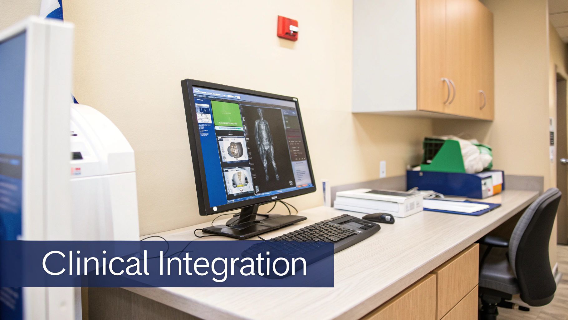
Making AI a Natural Part of the Clinical Workflow
Let’s be honest: a perfect AI model is completely useless if a clinician has to jump through hoops to use it. The real test of any AI medical imaging software isn't just accuracy; it's how invisibly it can weave itself into the fast-paced, high-stakes environment of a hospital.
This all comes down to interoperability. Your software has to speak the same language as the rest of the hospital's tech. In the world of radiology, that language is DICOM (Digital Imaging and Communications in Medicine). It’s the universal standard for everything from MRI scans to CT images, and supporting it isn't optional; it's the price of entry.
Connecting to the Hospital's Core Systems: PACS and RIS
Your AI tool can't live on an island. It needs to plug directly into the hospital's central nervous system, which means connecting with two key platforms:
PACS (Picture Archiving and Communication System): Think of this as the hospital's massive digital library for every medical image. Your AI needs to be able to seamlessly pull scans from the PACS for analysis and, just as importantly, push its findings back.
RIS (Radiology Information System): This is the logistical hub that handles patient scheduling, billing, and the radiologist's reports. Integrating here means your AI's insights can be automatically linked to the right patient file and reporting workflow.
Building these digital bridges is tricky. You're often connecting a modern AI application with legacy hospital systems. This is where leaning on experts in custom software development services really pays off. A good team makes sure that data flows securely and reliably, so clinicians aren't stuck toggling between different windows or applications.
Imagine this real-world scenario: a patient comes into the emergency department with a wrist injury. The X-ray is taken and sent to the hospital’s PACS. Your AI, which is constantly monitoring the PACS queue, automatically grabs the new study. It analyses the images and flags a potential scaphoid fracture – a tiny but critical injury that's notoriously easy to miss.
Within minutes, the AI sends its analysis back to the PACS, not as a clunky separate file, but as a new DICOM object. This could be an overlay that highlights the potential fracture right on the original X-ray. The radiologist opens the case in their usual viewing software and immediately sees the AI-generated alert, drawing their eye to the area of concern.
That’s the dream workflow. The AI isn't an extra step; it's an enhancement to an existing one. It acts like a second set of eyes, working quietly in the background to boost a clinician's confidence without ever disrupting their flow.
Choosing Your Deployment Model
A huge architectural decision is figuring out where your AI software will actually run. This choice impacts everything from security and speed to cost and scalability. You've essentially got three main paths, each with its own pros and cons.
| Deployment Model | Key Advantages | Best-Suited For |
|---|---|---|
| On-Premise | Gives the hospital maximum control over data security and privacy. Often has lower latency. | Large hospital networks with established IT infrastructure and very strict data governance rules. |
| Cloud-Based | Highly scalable, involves lower upfront hardware costs, and simplifies maintenance. | Startups and smaller diagnostic centres that need flexibility and want to avoid managing physical servers. |
| Hybrid | A mix of both worlds. Keeps sensitive patient data on-site while using the cloud for heavy-duty AI processing. | Organisations that want the security of on-premise with the computational power and flexibility of the cloud. |
There's no single right answer here. The best choice almost always depends on the client's budget, IT maturity, and security posture. Building a flexible architecture that can support more than one model is often the smartest long-term play.
Why MLOps Is Non-Negotiable for Long-Term Success
Getting your AI tool deployed isn't the end of the journey; it's the beginning. Medicine is always changing. New imaging equipment is introduced, patient demographics shift, and disease presentations evolve. An AI model that’s brilliant on launch day can slowly lose its edge over time. This decay in performance is a real phenomenon called model drift.
This is precisely where MLOps (Machine Learning Operations) becomes your safety net. MLOps is a discipline focused on making sure your AI models stay effective and reliable long after they've been deployed.
Think of it as a continuous quality control loop that includes:
Constant Monitoring: Keeping a close watch on your model's real-world performance to catch any dips in accuracy.
Automated Retraining: Having a system in place to periodically retrain the model on fresh, validated clinical data to keep it sharp.
Smart Version Control: Carefully managing different versions of your models so you can easily roll back to a stable version if an update causes unexpected issues.
Without a solid MLOps strategy, you're essentially shipping a static product that will inevitably become outdated and less trustworthy. It’s this commitment to ongoing performance that turns a good idea into a lasting, valuable clinical tool. This focus on long-term reliability is a cornerstone of our AI development services; it's how you build real trust with your clinical partners.
Navigating Healthcare Compliance and Security
When you're building medical software, brilliant innovation has to share the stage with security and privacy. These aren't just extra features you bolt on at the end; they're the absolute bedrock of your product. If you want clinicians and patients to trust your solution, you have to prove you can protect their most sensitive data. That means getting comfortable with the complex world of healthcare regulations.
For any AI medical imaging software development project here in Canada, the Personal Information Protection and Electronic Documents Act (PIPEDA) is the law of the land. It’s our equivalent of HIPAA in the U.S. and sets the rules for how organisations handle personal information. From the second you acquire patient data to the moment you securely delete it, every action is governed by these principles.
Building a Fortress Around Patient Data
Saying you’re compliant is one thing; proving it is another. It means baking specific technical and administrative safeguards into your software’s architecture right from the start.
Here’s what that looks like in practice:
End-to-End Encryption: This is non-negotiable. Data has to be encrypted both when it’s moving between systems (in transit) and when it’s sitting on a server (at rest). If someone manages to intercept it, it should be completely unreadable.
Robust Access Controls: You need to implement strict role-based access control (RBAC). A radiologist needs to see patient scans, but does the billing administrator? Absolutely not. RBAC ensures people can only see the information essential for their specific job.
Comprehensive Audit Trails: Your system must keep a detailed log of every single time someone interacts with patient data. Who looked at it? When? What did they do? These logs are crucial for accountability and for tracing the source of any potential breach.
For startups in this space, getting a handle on cybersecurity compliance frameworks like SOC 2 or ISO 27001 is a critical first step.
Demystifying Medical Device Classification
Beyond data privacy, you have to face another reality: your software will almost certainly be regulated as a medical device. Health Canada and the American FDA have specific rules for what they call Software as a Medical Device (SaMD). Your software will be assigned a classification, from low-risk Class I to high-risk Class III, based entirely on what it does and the potential for harm if it gets something wrong.
For example, an AI tool that just helps a radiologist prioritise their worklist might be seen as lower risk. But if your algorithm is making a direct diagnostic suggestion that will influence a patient's treatment? You can bet it will land in a much higher-risk category and face intense scrutiny.
The regulatory submission process is its own specialised field. It’s not for the faint of heart. You have to meticulously document everything – where your data came from, how you validated your model, your risk analysis, usability studies… all of it. Missing a single piece of evidence can mean long delays or even an outright rejection.
This is where finding a development partner who lives and breathes these regulations gives you a massive advantage. It helps you avoid incredibly costly missteps. It means that from the very first line of code, your software is built to meet the uncompromising standards of healthcare. For a closer look at these challenges, we dive deeper into AI in healthcare data privacy in Canada.
Getting Your AI into the Clinic: The Path to Adoption
Building a brilliant algorithm is one thing, but getting it into the hands of clinicians and seeing it used every day is the real measure of success. An AI tool that sits on a server, unused, is just a costly experiment. This is where your go-to-market strategy becomes just as critical as your model's architecture.
You can't do this alone. The key is to build genuine partnerships with the people on the ground – the hospitals, healthcare networks, and imaging centres. Think of these as collaborations, not just sales. These relationships are your direct line to invaluable feedback and can even lead to co-developing features that solve problems clinicians are actually facing.
Building for the Real World of Clinical Work
You absolutely have to put the user first. Before your team even thinks about writing code for a new feature, you need to live and breathe the clinical workflow it's supposed to improve. How do you confirm you've got it right? Through rigorous User Acceptance Testing (UAT).
This is where you put your software in front of the real end-users: radiologists, technologists, and even administrators. Let them run it through its paces in a setting that mimics their own.
Their feedback is everything. It will quickly show you where the friction is, what’s confusing, and what’s actually helpful. The goal is to make your software feel like a seamless part of their existing toolkit, not another clunky system they have to fight with.
At the end of the day, adoption comes down to trust. Doctors and medical staff need to be confident that your AI is reliable, that they can understand its outputs, and that it genuinely makes their incredibly demanding jobs a bit easier. You don't get that trust for free; you earn it with transparency and solid validation.
More Than Just Installation: Training and Support
Dropping off the software and waving goodbye is a recipe for failure. A comprehensive training program is essential to get staff comfortable and help them move past any initial scepticism about new technology.
A training plan that actually works usually includes:
Hands-on Sessions: Let clinicians work with the software using real, anonymised cases. This builds muscle memory and confidence.
Straightforward Guides: Create documentation that explains not just how to click the buttons, but also gives a basic rundown of what the AI is doing and why.
A Real Human to Call: Provide a direct line to product specialists who can answer both technical and clinical questions without a long wait.
We see ourselves as more than just developers; we're partners in bringing new ideas to life. Our work in both AI development services and custom software development services is built on this idea of working together. To see what drives our commitment to improving healthcare, you can learn more about our team on our about us page.
Got Questions? We've Got Answers
If you're delving into AI medical imaging, you've probably got a lot of questions. It's a complex field, after all. Below, I’ve answered some of the most common queries we hear from our clients, breaking down what you really need to know.
How Long Does It Take to Build a Custom AI Medical Imaging Solution?
That's the million-dollar question, isn't it? The honest answer is: it depends entirely on the scope. A focused proof-of-concept (PoC) to test a specific hypothesis might come together in 6-9 months. But if you're building a fully-featured, clinically validated product that needs regulatory approval, you should plan for 18-24 months, and sometimes even longer.
The timeline really hinges on a few key things. The availability and quality of your training data is a huge one. Then there's the complexity of the AI model itself and how deeply it needs to integrate with hospital systems like PACS and EMRs. And of course, the regulatory dance with bodies like Health Canada or the FDA always adds a significant chunk of time.
What Are the Biggest Hurdles in This Field?
From my experience, it boils down to three major challenges that can make or break a project. First, it’s all about data accessibility and quality. Getting your hands on large, diverse, and perfectly annotated datasets, while staying on the right side of privacy laws like PIPEDA, is a monumental task right from the start.
Next up is clinical integration. An algorithm can be brilliant, but if it doesn't seamlessly fit into a radiologist's existing workflow and talk to their PACS/RIS systems, it’s dead in the water. Usability is just as important as accuracy. The final mountain to climb is regulatory compliance; navigating the complex approval process for medical device software is non-negotiable to ensure the tool is safe and effective for patient care.
A project's success isn't just about technical brilliance. It's about mastering data, integration, and regulation. Drop the ball on any one of these, and even the most groundbreaking AI model will fail to make an impact.
How Can You Be Sure the AI Model Is Accurate and Reliable?
Building trust in an AI model is a step-by-step process, not a single event. It all starts with the data – if you put garbage in, you'll get garbage out. So, high-quality, expertly annotated data is the absolute foundation. From there, we move into rigorous technical validation, where we relentlessly test the model against data it's never seen before, measuring things like sensitivity, specificity, and overall accuracy.
But the real acid test is clinical validation. This is where we put the model to work with practising radiologists in real-world scenarios. It’s the only way to prove it actually provides diagnostic value and doesn't just look good on paper. Once deployed, our approach to AI development services involves continuous MLOps and monitoring to make sure performance stays sharp and reliable for the long haul.
At Cleffex, our focus is on turning tough healthcare problems into software solutions that are reliable, compliant, and genuinely useful in a clinical setting. If you’re ready to build the next generation of medical imaging tools, reach out and let's talk about your project.


